Penn State films Zika and chikungunya RNA live in lab cells
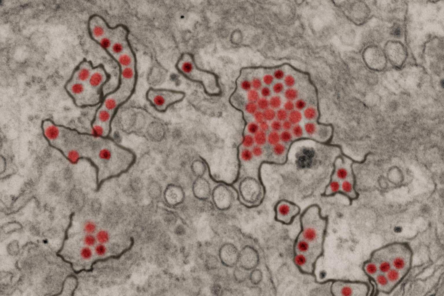
Inside a cell, a virus hides in plain sight. For Zika and chikungunya, two mosquito-borne viruses whose genetic material is RNA, scientists have largely been left in the dark about what happens after infection, because few tools can follow the virus as it moves through a living cell. At Penn State, a team led by Joyce Jose, associate professor of biochemistry and molecular biology, has now filmed the viral RNA of both viruses moving inside live infected cells in the laboratory.
The key is a tag called Mango-II, a short sequence of DNA or RNA inserted directly into the virus's genome. It folds into shapes on the single-stranded viral RNA and binds to a stain that glows bright green under the microscope. When the virus enters its double-stranded replication stage, the glow goes dark. Placing the tag was the hard part: the genome is so compact that almost all of it is essential, and the usual insertion site would stop the virus from replicating. The team put the tag in the protein-coding region instead, something that had not been done before to visualize viral RNA. According to the team, the tag does not affect the virus's life cycle.
The two viruses moved differently. Zika's RNA is anchored to the endoplasmic reticulum, the cell's main manufacturing and transport network, whereas chikungunya's RNA travels more widely and more rapidly along the plasma membrane. Jose compares the older tools to a photograph: electron microscopy captures fixed images and loses the moment-to-moment interactions. Live imaging, she says, is "the difference between a photograph and a video." The team says the movement patterns matched what earlier published studies had described.
The same tag also served as a fishing line. Once the researchers extracted the tagged viral RNA from infected cells, they identified, after comparing against two controls, more than 200 host proteins that interacted with Zika virus with high confidence, and a smaller set for chikungunya. 132 host factors are shared by the two viruses. These are the cell proteins the viruses rely on to survive, and each one is a possible point where a drug could interrupt the infection.
For patients, the gain is indirect and far off. Jose notes there are currently no good antiviral treatments for either infection. The study does not announce a drug or a date, and its experiments were run in commercially available human cells in the lab. What it gives antiviral researchers is a map of who, what, where and when the virus uses the host cell, drawn from a method that leaves the infection intact. Jose says it could be applied to dengue, hepatitis C and rhinovirus. The team, by her account, spent eight to nine years building toward this live imaging.
Comments
Loading the thread…
Sign in to leave a comment. Sign in