In the lab, antibody access to cancer vesicles varies widely
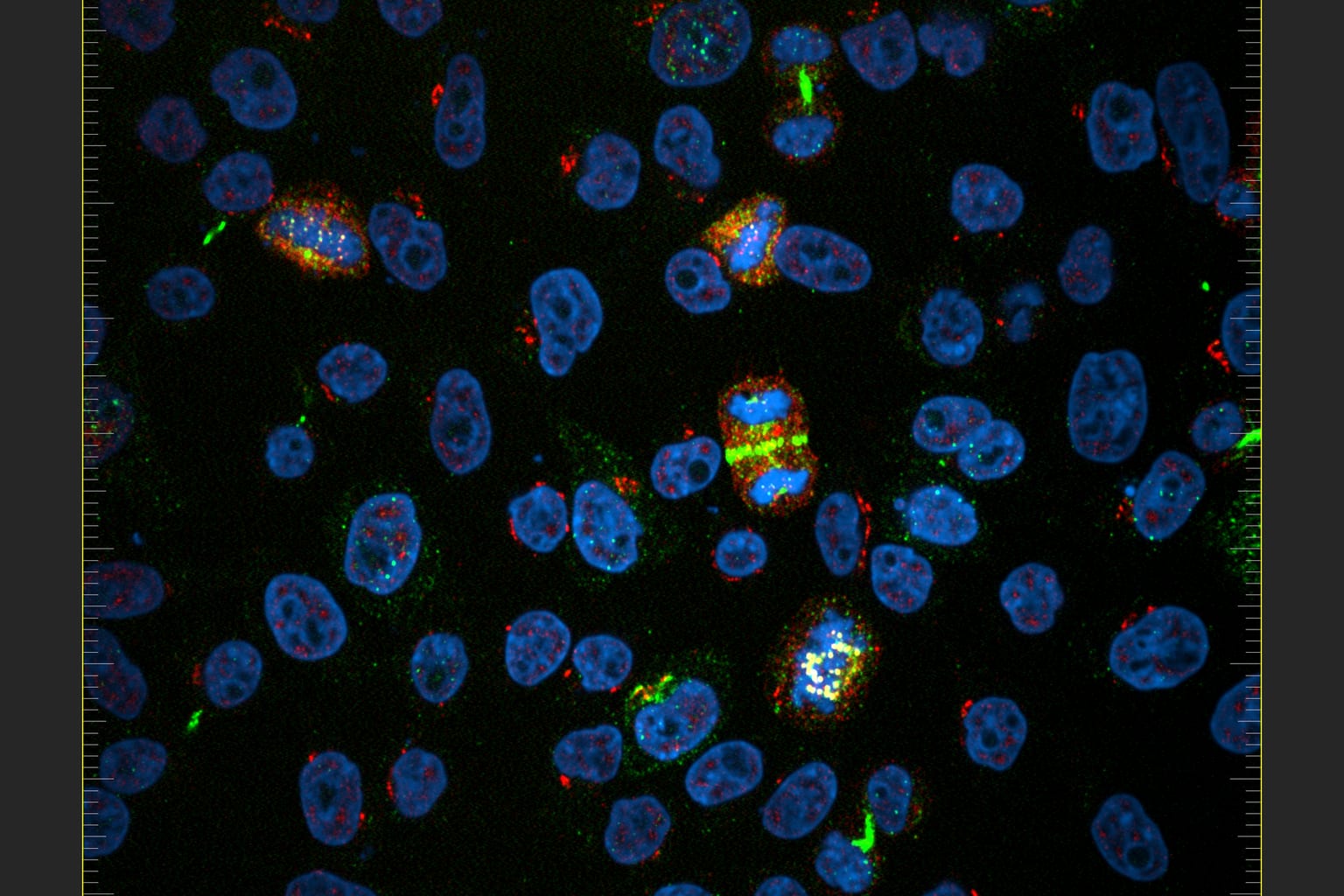
In a drop of liquid, an extremely fine probe slides across a surface and builds images at the nanoscale, and in the frames a few antibodies land, stay for a moment, or slip away. That is the view Kanazawa University researchers used to watch single cancer-linked vesicles, the tiny packages cells release, in real time.
Cancer cells can switch off the immune system in several ways, and one of them runs through PD-L1, a protein that acts as a brake on immune cells able to attack a tumor. Medicines that block this interaction have transformed the treatment of some cancers, but patients respond very differently. Standard tests mostly count how much PD-L1 is present. Yet a protein can be there and still be partly hidden by neighbouring molecules, or placed so that an antibody struggles to reach it. Researchers call this physical availability accessibility, and until now it was hard to measure on individual vesicles.
The team used high-speed atomic force microscopy, which needs neither fluorescent labels nor chemical fixation, so the antibodies could be followed as they bound. They compared vesicles from two breast cancer cell lines. MCF7 cells carry relatively little PD-L1, while MDA-MB-231 cells, a model of triple-negative breast cancer, express much more. Antibodies behaved very differently on the two groups: on high-PD-L1 vesicles they often docked and stayed, and on low-PD-L1 vesicles they moved along the surface and vanished from view. Over 80% of high-PD-L1 vesicles showed persistent association, while over 90% of low-PD-L1 vesicles showed no detectable stable interaction. A second antibody, aimed at CD63, a marker common on vesicles, gave broadly similar patterns, which suggests the differences came from PD-L1 itself and not from a general stickiness of some vesicles.
The method also worked on material from a patient. Vesicles from cells grown in the laboratory from one patient's triple-negative tumor showed detectable antibody interactions on 93.5% of vesicles, with binding duration falling between the two cell-line models. The authors stress the limits: one person, cells grown in a dish, and no evidence yet that the method predicts whether a patient will respond to immunotherapy. The team calls its classification, based on how often and how long antibodies bind, the Checkpoint Ligand Interface Phenotype, or CLIP framework, and says it remains an experimental tool rather than a diagnostic one.
For now, the change is in what researchers can see. Averages across large groups of vesicles can hide the variation that this imaging reveals, so studies of tumor vesicles could gain a finer picture of who is reachable by treatment. Doctors and patients are not affected yet: the work needs further research before any clinical use, and the team gives no timeline for that step.
Comments
Loading the thread…
Sign in to leave a comment. Sign in